

COLUMBUS, OH (OSU) - From 2D to 3D, doctors are turning medical imaging, like MRI and CT scans, into 3-dimensional models to better treat and personalize care for patients battling jaw cancer. Karina Chung shares how state-of-the-art 3D printing technology is making a difference for doctors and patients.

A routine trip to the dentist. That was when Rick Turner was first alerted that something wasn't right.
"The hygienist said, uh, Rick. I don't like the way the tissue looks by your molars in the upper right," said Rick Turner, patient.

Testing revealed cancer in Rick's upper jawbone. And invasive and complex surgery was the best option to save his life.
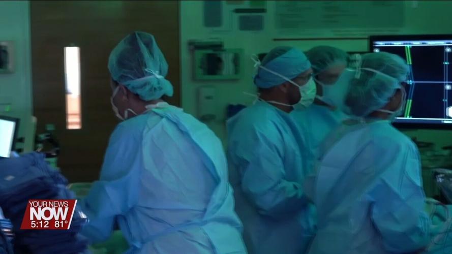
"We want to make sure these patients can talk, can chew, can eat, can swallow, can breathe normally, and function normally as much as possible," stated Dr. Kyle Vankoevering, MD.
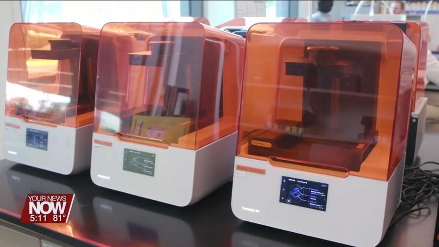
So Dr. Kyle Vankoevering and his team at the Ohio State University Comprehensive Cancer Center—James Cancer Hospital and Solove Research Institute pioneered a new tool to aid doctors in jaw reconstruction surgery.

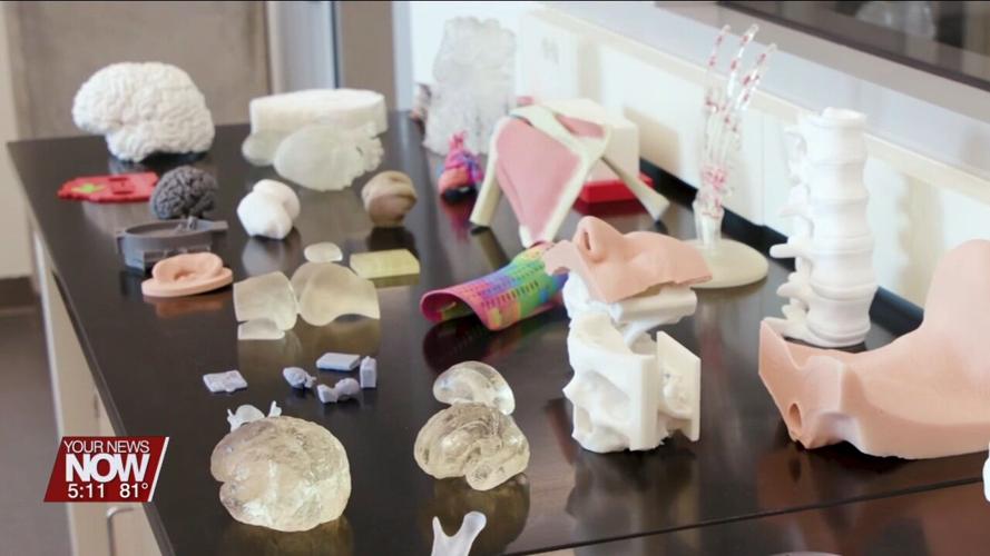
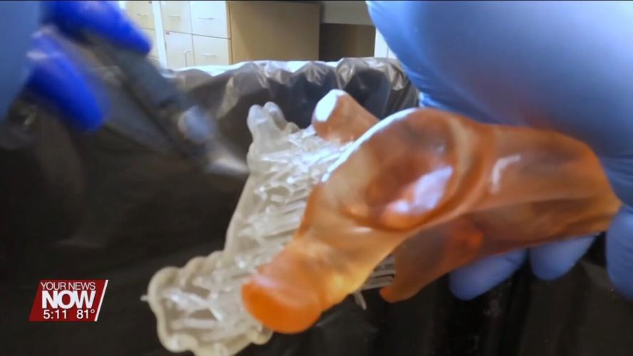
Using MRI and CT scans, doctors 3D print an exact replica of their patient's jaw, helping to guide critical decisions in the OR.
"We can actually bring a real-life template into the operating room that gives us a lot more precision on where to make the cuts and how to align things afterward," explained Dr. Vankoevering.
The model is also used to custom tailor and shape new bone to replace what was removed, which is often taken from a patient's arm or leg.
"The bone grafts we bring in heal better, have less likelihood of fracture and lower rates of infection in the long run, which is going to improve the outcomes for our patients," added Dr. Vankoevering.
This life-saving procedure removed Rick's cancer. He's thankful for the 3D technology, crediting it to how he looks and feels today.
"I'm sure it would be difficult to get exactly the same results without having the tool to help," said Turner.